Hemolyzed Samples: Recognizing and Preventing Sample Collection Errors
It was a case that puzzled the evening shift. A 68-year-old male in the cardiac unit had a routine metabolic panel drawn. The lab immediately called a critical result: his potassium was 10.0 mmol/L, a level high enough to cause a life-threatening arrhythmia. The clinical team prepared for an emergency intervention. But one experienced clinical laboratory director paused. The patient’s EKG was stable, and he showed none of the classic signs of hyperkalemia. He looked at the note from the lab: “Sample is moderately hemolyzed.” This was not a true critical value; it was a red herring a false clue created by a collection error that almost led to a dangerous and unnecessary treatment.
This story is a powerful reminder that hemolysis is more than just an inconvenience; it’s a direct threat to patient safety.
This blog post is designed for the meticulous healthcare professional dedicated to diagnostic accuracy. We will dissect the causes of hemolysis, explore its profound impact on common lab tests, and provide evidence-based best practices to help you protect the integrity of every sample and prevent these critical errors.
Understanding Hemolysis: A Breakdown of the Basics
To defeat the enemy, you must know it. Let’s explore the fundamentals of hemolysis.
What is Hemolysis and Why Does It Matter in Lab Testing
Hemolysis is the breakdown of red blood cells (erythrocytes), which causes their internal contents to spill into the surrounding serum or plasma. This is a critical issue because it fundamentally contaminates the sample. Laboratory analyzers are meticulously calibrated to measure the chemistry of clean plasma/serum. When that fluid is flooded with intracellular components like potassium and enzymes, the results become clinically unreliable and potentially misleading.
Hemolysis vs. Intravascular Hemolysis: Know the Difference
A critical skill is distinguishing in vitro hemolysis (from collection) from in vivo hemolysis (occurring in the body). In vivo hemolysis is a sign of a true medical condition and is a diagnostic clue itself. Lab professionals and physicians work together, using other lab results and the patient’s clinical presentation, to make this crucial distinction.
The Science Behind Hemolysis: What Happens to Blood Cells?
A red blood cells membrane is designed for flexibility, not for withstanding intense physical force. During a traumatic blood draw or rough handling, this membrane can rupture. The release of hemoglobin gives the sample its characteristic pink or red color, but the real danger lies in the invisible components that leak out especially the high concentration of potassium inside the cell.
Common Signs That a Sample Is Hemolyzed
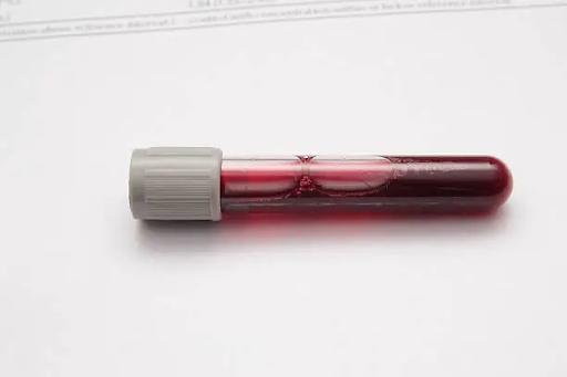
Visual inspection is the first line of defense. Serum or plasma should be a clear, straw-yellow color. Hemolysis will turn it pink (slight), red (moderate), or dark brown (gross). Modern analyzers also provide a quantitative “hemolysis index,” but visual recognition remains a vital skill for both lab and collection staff. The evidence is clear: hemolysis is a major problem. Hemolysis is one of the leading causes of specimen rejection in clinical labs. So, where are these errors originating?
Causes of Hemolysis: From Collection to Handling
The overwhelming majority of hemolysis is in vitro—meaning it happens outside the body during or after collection. Let’s trace the journey of a sample to identify the common failure points.
Poor Phlebotomy Techniques That Lead to Hemolysis
- Needle Selection: Using a needle with too small a bore (e.g., >22 gauge) for venipuncture creates shear stress that shreds red blood cells.
- Tourniquet Time: Application for over one minute leads to hemoconcentration and increases cellular fragility.
- Collection Method: Using a syringe requires a delicate touch. Pulling the plunger too fast or forcefully transferring the blood into a vacuum tube are common causes of cell rupture.
- Site Selection: Drawing from an IV catheter without a proper flush and discard can introduce fluids and pressure changes that lead to hemolysis.
How Transportation and Storage Affect Sample Integrity
- Mixing Technique: Tubes with additives must be mixed by gentle inversion 5-10 times. Vigorous shaking is one of the most common handling errors.
- Temperature and Storage: Exposure to extreme heat or cold can lyse cells.
- Transport Method: Pneumatic tube systems, if not well-maintained, can subject samples to extreme G-forces, causing what’s known as “roller-coaster hemolysis.”
Now that we’ve identified the causes, let’s examine the specific and dangerous consequences of these errors on patient test results.
Impact of Hemolysis on Laboratory Test Results
Which Blood Tests Are Most Affected by Hemolysis?
The tests most impacted are those for analytes with high intracellular concentrations.
- Potassium (K+): The most dangerous interference. Falsely high potassium can lead to incorrect emergency treatment.
- LDH and AST: These enzymes are abundant in red blood cells. False elevations can mimic signs of liver or heart injury.
- Magnesium and Phosphate: Also falsely elevated.
- Troponin: While not inside the red cell, some troponin assays can be negatively interfered with by components released during hemolysis, potentially masking a true heart attack.
Examples of Lab Values Altered by Hemolyzed Samples
To understand the real-world impact, consider this scenario:
A patient’s reference potassium level is within 3.6 – 4.2 mmol/L (a healthy, normal value). A moderately hemolyzed sample could falsely report this value as 6.5 mmol/L, a critical result that suggests a life-threatening condition.
A patient’s reference AST level within 8 to 48 U/L. (normal). Hemolysis could cause this to be reported as 150 U/L, falsely suggesting acute liver injurY. These are not just numbers on a page; they are false signals that can lead to incorrect diagnoses and unnecessary, potentially harmful treatments.
A patient’s reference potassium level is within 3.6 – 4.2 mmol/L (a healthy, normal value). A moderately hemolyzed sample could falsely report this value as 6.5 mmol/L, a critical result that suggests a life-threatening condition.
How Hemolysis Can Lead to Misdiagnosis or Delayed Treatment
The case study at the beginning illustrates the risk of misdiagnosis. More commonly, hemolysis leads to significant delays. A pre-surgical screen is rejected, and a necessary surgery is postponed. An ER result is delayed, extending the time a patient waits for a diagnosis for critical conditions like a heart attack or sepsis. These delays add up, impacting patient care, length of hospital stay, and overall work efficiency.
So, a compromised sample has arrived in the lab. What is the standard protocol, and what happens next?
Dealing with Hemolyzed Samples in Clinical Practice
When a sample is identified as hemolyzed, the lab initiates a strict, systematic process to ensure patient safety.
What Happens When a Sample Is Rejected Due to Hemolysis?
- Preventing hemolysis is rooted in protocol, precision, and continuous education.
- Identification: The lab technologist identifies hemolysis either visually or via an automated flag from the analyzer.
- Documentation: The sample is documented as rejected in the Laboratory Information System (LIS), and the reason (e.g., “moderate hemolysis”) is recorded. The compromised result is suppressed.
- Communication: The technologist contacts the collecting unit (e.g., the nursing floor, ER, or outpatient clinic) to inform them of the rejection.
- Recollection Request: A request for a new sample is made. This is the “redraw”—the crucial step that closes the loop on the error.
Can Hemolyzed Samples Be Used for Any Tests?
This is a frequent and important question. The answer is nuanced: it depends on the lab’s policy and the degree of hemolysis.
- For slight hemolysis, a lab might release results for tests known to be unaffected (e.g., glucose, creatinine, sodium).
- However, all results for highly affected analytes (like potassium, LDH, AST) will be canceled.
- For moderate to gross hemolysis, most facilities will reject the entire sample, as the integrity of all results is questionable. The risk of releasing a misleading result is too high.
Recollection Guidelines: When and Why It’s Necessary
A recollection is necessary whenever a test result critical to a clinical decision is compromised.
- Why it’s necessary: The primary reason is patient safety. Acting on a falsely elevated potassium level is dangerous. Failing to act on a falsely lowered troponin level can be fatal. A redraw is the only way to ensure the clinical team has accurate data.
- When it’s necessary: As soon as the lab communicates the rejection. The goal of the recollection is not just to obtain a new sample, but to use it as a learning opportunity to review the initial collection technique and make corrections to prevent a repeat error.
The cycle of redraws is frustrating, but it is not inevitable. Here are the evidence-based strategies to get it right the first time.
Preventing Hemolysis: Best Practices for Accurate Results
Prevention is rooted in protocol, precision, and continuous education.
Step-by-Step Guide to Proper Blood Collection Techniques
- Use a 21-22G needle for routine draws.
- Ensure tourniquet time is under 60 seconds.
- Allow vacuum tubes to fill completely and naturally.
- If using a syringe, remove the needle before transferring blood, and let the tube’s vacuum draw the blood in gently.
- Invert don’t shake: Gently invert tubes with additives 5-10 times to ensure proper mixing without damaging cells.
Tips for Handling and Transporting Samples Without Damage
A perfect draw can be ruined on the journey to the lab.
- The Golden Rule: Invert, Don’t Shake. This is the single most important handling tip. For tubes with additives, gentle inversion 8-10 times is sufficient to mix the sample. Shaking creates intense mechanical trauma.
- Maintain Temperature Stability: Avoid leaving samples in extreme temperatures, like a hot car dashboard or directly on ice (unless specifically required for the test).
- Secure and Upright: Keep tubes upright in a rack during transport. This minimizes agitation and, for serum tubes, allows for proper clot formation.
Quality Control and Lab Protocols
For managers and senior lab staff, a systems-level view is essential for quality improvement.
Lab SOPs and Continuous Education
Facilities must have clear Standard Operating Procedures (SOPs) for handling and documenting hemolyzed samples.
More importantly, tracking hemolysis rates by department can identify areas that need targeted re-education.
Providing non-punitive, constructive feedback and hands-on training refreshers are the most effective ways to reduce institutional rates.
Conclusion: Upholding a Standard of Excellence
The story of the misidentified critical potassium level is not an isolated incident; it’s a scenario that plays out in healthcare facilities every day. It underscores a fundamental truth: the integrity of our most advanced diagnostic tools is entirely dependent on the integrity of the sample we provide. Preventing hemolysis is not a minor detail; it is a professional obligation. It is a direct reflection of our commitment to patient safety, accuracy, and efficiency.
Share Your Story, Help Our Community
Do you have a “redraw story” you’ll never forget—a time when a tricky situation taught you a valuable lesson. Let us know


latest video

news via inbox
From regulatory updates to cutting-edge technology, we've got you covered.